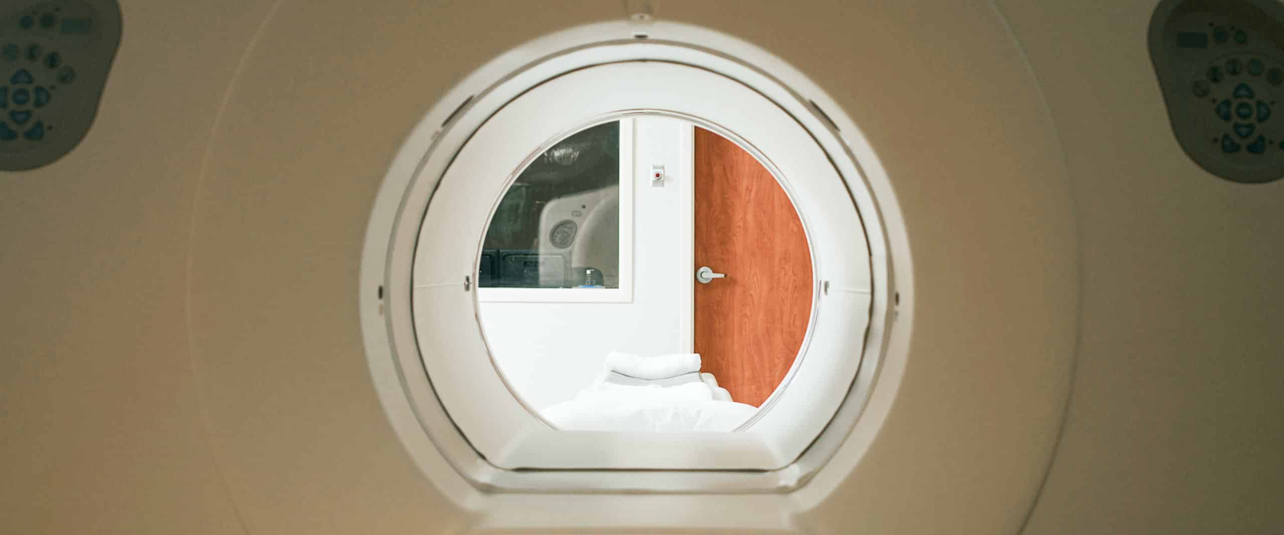
To ensure you receive the best possible care, it’s essential for us to gain a detailed insight into your health. At our state-of-the-art Imaging Center, our dedicated staff will guide you through each step, explaining the process and ensuring your comfort. We conduct extensive tests not just to diagnose current concerns, but also to identify and understand any potential future issues.
Accreditation is a testament to our commitment to excellence. When you undergo an examination at Cigarroa Clinic, rest assured that we adhere to the latest standards of care. We are proud to be accredited by the Intersocietal Accreditation Commission for Nuclear Medicine, Nuclear Cardiology, and PET. Additionally, we have received accreditation from the American College of Radiology for Computed Tomography (CT) imaging in adult patients.

The staff will attach a small wearable monitor to your chest to evaluate your heart rhythm.
• Provides a ‘real-life’ view as it checks and records your heart rate while you go about your daily activities
• Evaluates symptoms associated with dizziness or shortness of breath
• It is a non-invasive test (electrodes are placed on the chest)
Radiographic image to see inside your body, this painless procedure helps give us an accurate picture of any issues affecting your soft tissue or bones.
• Tested areas often include chest, abdomen, hands, and feet
• Takes 10 minutes
• Checks for conditions such as abdominal pain, trauma, joint pain and arthritis
• Non-invasive
This important study creates ultrasound pictures of your heart and evaluates the heart muscle and valves. It can detect poorly functioning valves, extra fluild around the heart an look at the valves between the upper and lower chambers.
• Procedure to measure blood flow and valve function
• Produces an ejection fraction—a percentage that indicates how much blood is pumped out of the left bottom chamber (ventricle)
• Takes 20 minutes
• Non-invasive
An ultrasound uses high-frequency sound waves to capture “live” moving images of your organs from inside your body.
• Commonly studies include the neck, thyroid, and abdomen
• Takes 20 minutes
• Non-invasive


A CT provides more detail than x-rays by using computer-processing to create cross sectional images or slices of your internal organs and blood vessels.
CTA uses CT technology along with an injection of dye to see more detail of one or more areas of interest. The procedure allows the visualization of a 3D images of the heart and vessels as they move. The procedure is minimally invasive due to placement of an intravenous line (IV) for medication administration.
• Used to check for heart disease or any other blocked blood vessel
• A CT scan takes 15 minutes and CTA scan takes 20 minutes
• Areas frequently scanned are the head, neck, chest, and abdomen
• The thyroid scan includes the uptake of the dye to see the thyroid at work
A highly specialized area of medicine where small amounts of radioactive dye is used to examine organ function and structure. It means one can see a clear view of your tissue and evaluate the vessels for blood clots or damage. The discipline is highly regulated due to the use of the dye. The procedure is minimally invasive due to IV line placement.
The following test may be ordered:
• Nuclear stress test, to understand your heart muscle and blood flow
• Exercise thallium, which combines heart activity with exercise to examine how your heart is performing during stress
• MUGA scan assesses your heart’s muscle strength of the lower chambers
• PET scans uses dye to evaluate the heart for disease, poor blood flow, muscle function, enlarged heart, or other conditions (including cancer).
A probe is uses sound waves to measure the amount of blood flow through arteries and veins.
• Focuses on your brain function, carotid arteries, and/or arms and legs
• Usually takes around 30-45 minutes
• Non-invasive
